Abdominal Ultrasound

Dr. Sotiria Bedreli offers specialized medical services in the field of ultrasound.

Ultrasound of the Liver, Gallbladder & Bile duct, Spleen, Pancreas, Intestine, Abdominal Vessels

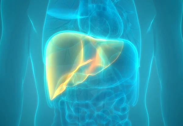
In our private practice in Alexandroupolis we perform ultrasound scans of all organs of the gastrointestinal system, including the liver, gallbladder and bile ducts, spleen, pancreas, intestine, as well as abdominal vessels. The clinic is equipped with a high-end Canon ultrasound system.

Gastroenterologist-hepatologist Sotiria Bedreli is a certified ultrasonographer in both Greece and Germany and has many years of experience in evaluating all abdominal organs.

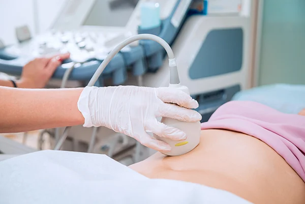
Abdominal ultrasound is a safe and painless examination method. Its performance is necessary in the context of investigating and evaluating any type of pain or symptomatology in the abdominal area, but also in the context of a preventive check-up.

Intestinal Ultrasound (IUS)

Intestinal Ultrasound (IUS) is, in the hands of an experienced examiner and using a modern ultrasound machine, an excellent tool for the diagnosis and monitoring of intestinal pathologies. For example, in inflammatory bowel diseases, such as ulcerative colitis and Crohn’s disease, it makes it possible to regularly, quickly and painlessly monitor the disease and its complications, without special patient preparation and without repeated endoscopies.

With intestinal ultrasound, it is possible to diagnose a wide range of intestinal diseases, such as diverticulitis, appendicitis, intestinal obstruction, as well as intestinal or stomach tumors.

Contrast-Enhanced UltraSound (CEUS)

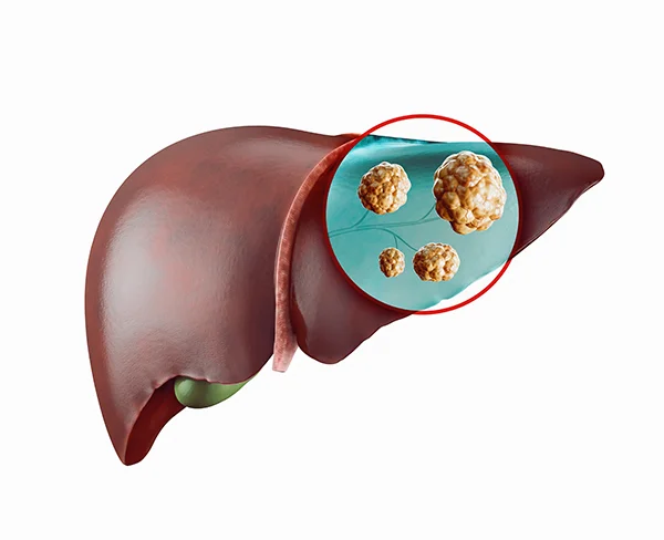
Contrast-enhanced ultrasound (CEUS) is a pioneering method for examining and identifying lesions, especially in the liver. It can identify a lesion as benign or malignant, thus avoiding unnecessary biopsy. It is also possible to further identify a liver lesion based on its vascularization (for example, hemangiomas, focal nodular hyperplasia, adenomas, regenerative liver nodules), often making diagnosis possible within the framework of a single ultrasound.

Elastography of the liver

At the hepatology practice of Sotiria Bedreli, in Alexandroupoli, it is possible to perform liver elastography, a modern method of examining the degree of fibrosis-scarring of the liver. In patients with chronic liver diseases of all forms (e.g. fatty infiltration, chronic hepatitis, alcoholic liver disease, autoimmune diseases), the severity of the disease can be assessed by measuring the degree of liver fibrosis, i.e. the elasticity of the organ, and even without having a biopsy.

Liver Elastography makes it possible to regularly monitor the course and treatment of chronic liver disease, as well as to evaluate the stage of the disease from the early stages of fibrosis to those of cirrhosis.

ATI (ATtenuation Imaging) of the liver

Fatty liver disease (steatosis), with or without inflammation (steatohepatitis), is the most common liver disease with a rapid increase in recent decades. It is a manifestation of metabolic syndrome and is associated with increased morbidity and mortality.

In our clinic, the degree of fatty infiltration is measured using the innovative ATI (ATenuation Imaging) method, which is a fast, painless and bloodless method of quantitatively measuring fatty infiltration of the liver. This avoids the invasive biopsy, allowing for regular repetition and immediate control of the result and success of a treatment.

The ATI method can be combined with the elastography method for the simultaneous analysis of the degree of fibrosis of the liver parenchyma due to fatty infiltration.

Gastroenterologist-hepatologist Sotiria Bedreli is an accredited sonographer, with excellent specialization in ultrasound of the organs of the gastrointestinal system.